
Anatomy of back muscles Diagram Quizlet
Back Muscles w/ pictures 18 terms Jenna_Carey1 Preview parts of the brain 33 terms kmd6gu Preview Muscle Labeling - Upper Leg 22 terms MonaL18 Preview Layers of the wall of the digestive tract 10 terms abreejtheriot Preview Muscle Labeling - Lower Leg

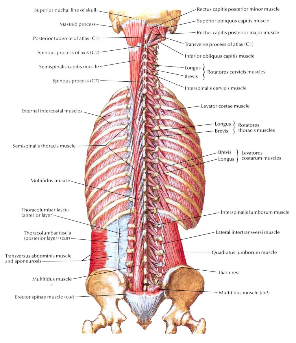
Intrinsic Back Muscles Anatomy of the Torso Medical Library
Labeling Exercises: Crossword Puzzles: Flashcards: Concentration: Internet Activities: Chapter Weblinks: Feedback Help Center: Human Anatomy, 6/e. Kent Van De Graaff, Weber State University. Muscular System. Labeling Exercises. Muscles-Anterior View 1 Muscles-Anterior View 2 Muscles- Anterior View 3

Human Anatomy Back Muscle diagram, Muscle anatomy, Lower back muscles
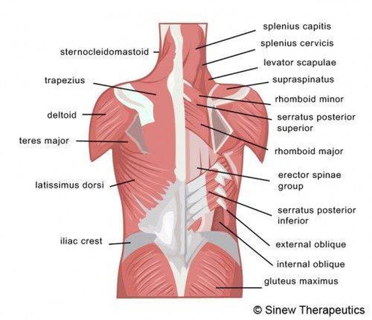
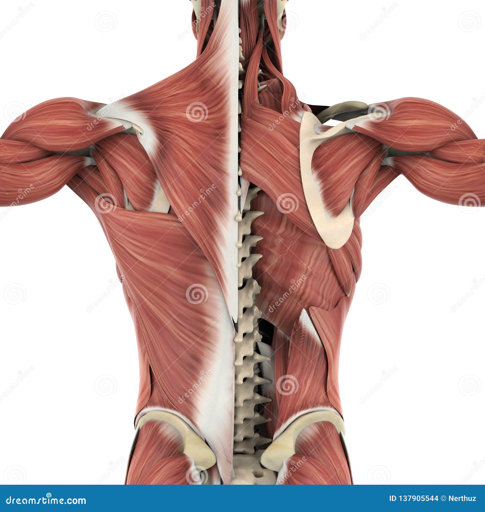
Muscles of the Back Image of the muscles of the back, labeled for reference and study.

Muscles of the Back Labeling Diagram Quizlet
Your back consists of a complex array of bones, discs, nerves, joints, and muscles. The muscles of your back support your spine, attach your pelvis and shoulders to your trunk, and provide mobility and stability to your trunk and spine. The anatomy of your back muscles can be complex. There are several different layers of muscles in your back.

Thoracic Mobility Impulse Chiro & Natural Therapies
The muscles of the back categorize into three groups. The intrinsic or deep muscles are those muscles that fuse with the vertebral column. The second group is the superficial muscles, which help with shoulder and neck movements. The final group is the intermediate muscles, which help with the movement of the thoracic cage.

Back Muscles Diagram Quizlet
What are your back muscles? Your back has many different muscles. Some muscles support your spine and trunk. Others help you move your body, stand up straight and assist with breathing. Because your back muscles support so much of your weight and are responsible for so many movements, injuries to these muscles are common.
Muscles of Back Deep Layers Biological Science Picture Directory
A back muscle that pulls the arm down and back. It is responsible for extension,adduction, and (medial) internal rotation of the shoulder joint. It also helps in extension and lateral flexion of the lumbar spine. The name means "widest of the back." This muscle supports the arm when it is moved above the head.

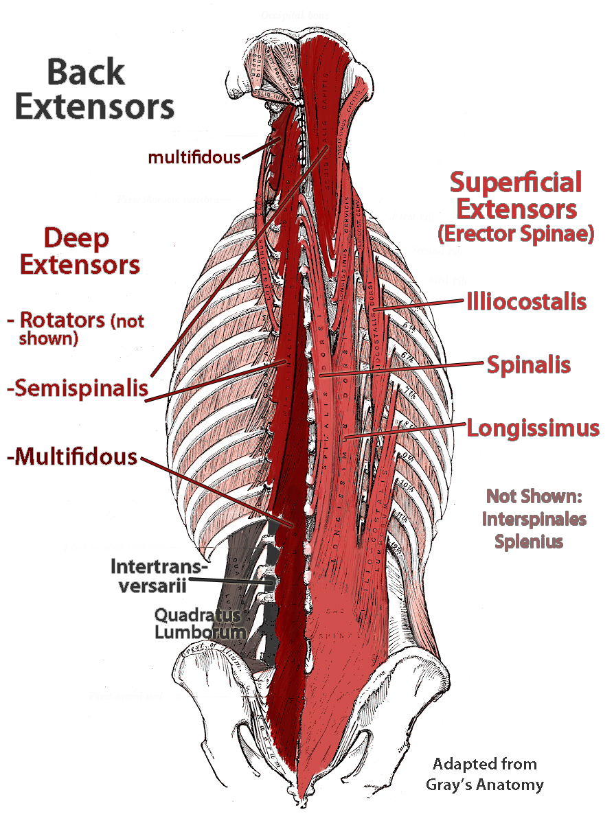
Deep muscles of the back labeling (erector spinae only) Diagram Quizlet
Erector spinae: three groups ("I long for spinach"), lateral to medial: illiocostalis: lateral. longissimus: in the middle. spinalis: medial. Transversospinales: three groups, from superficial to deep: semispinalis. multifidus. rotares. Learn all about the muscles of the back in this 3D video anatomy tutorial.

Back Muscle Diagrams 101 Diagrams
muscles of the back labeling by sofiaa17 1,042 plays 22 questions About a minute English 22p More 2 too few (you: not rated) Tries Unlimited [?] Last Played February 22, 2022 - 12:00 am There is a printable worksheet available for download here so you can take the quiz with pen and paper. Remaining 0 Correct 0 Wrong 0 Press play! 0% 0:00.0

Diagram Of Hip.and Back.muscles qwlearn
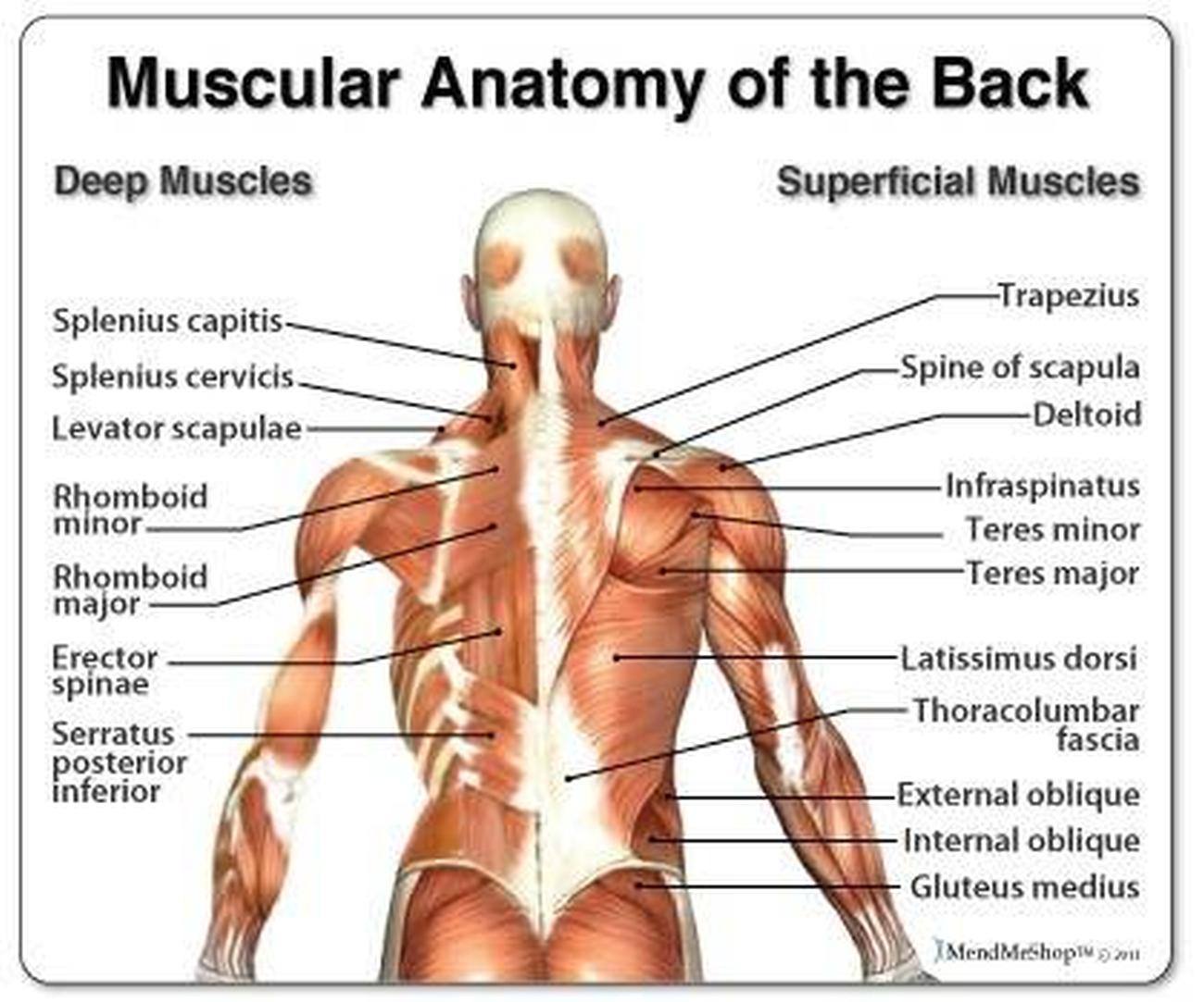
The muscles of the back are a group of strong, paired muscles that lie on the posterior aspect of the trunk. They provide movements of the spine, stability to the trunk, as well as the coordination between the movements of the limbs and trunk. The back muscles are divided into two large groups:
Pictures Of Back Muscles
The muscles of the back can be arranged into 3 categories based on their location: superficial back muscles, intermediate back muscles and intrinsic back muscles.The intrinsic muscles are named as such because their embryological development begins in the back, oppose to the superficial and intermediate back muscles which develop elsewhere and are therefore classed as extrinsic muscles.
deep muscles of lower back Biological Science Picture Directory
The back is found posteriorly and includes the vertebral column, the muscles that support the back and the spinal cord. The vertebral column consists of 33 vertebrae which can be split up into 5 continuous sections. Each section is functionally different and is specialised for either weight-bearing, movement, protection and/or posture.

Back Muscle Diagram exatin.info
Anatomy of the back: spine and back muscles Author: Jana Vasković MD • Reviewer: Nicola McLaren MSc Last reviewed: November 03, 2023 Reading time: 14 minutes Recommended video: Superficial back muscles [17:28] Attachments, innervation and functions of the superficial muscles of the back. Back anatomy
Pictures Of Back Muscles
Latissimus Dorsi Your latissimus dorsi, or lats, are the largest individual muscles in your upper back. They run down the sides of your torso and, when developed through resistance training,.

Muscles Labeled Front And Back Human Anatomy Body
Study with Quizlet and memorize flashcards containing terms like Longissimus Capitis, Splenius Capitis, Serratus Posterior Superior and more.
Muscle Chart Back Understanding Low Back Pain Anatomical Chart The Physio Shop
This online quiz is called Back muscles. It was created by member LilStride and has 13 questions.
